Below is a small preview from the upcoming book.

Photomicrograph of a neuron’s cell body (top, center) and its dendrites radiating out of it, obtained with a scanning electron microscope.
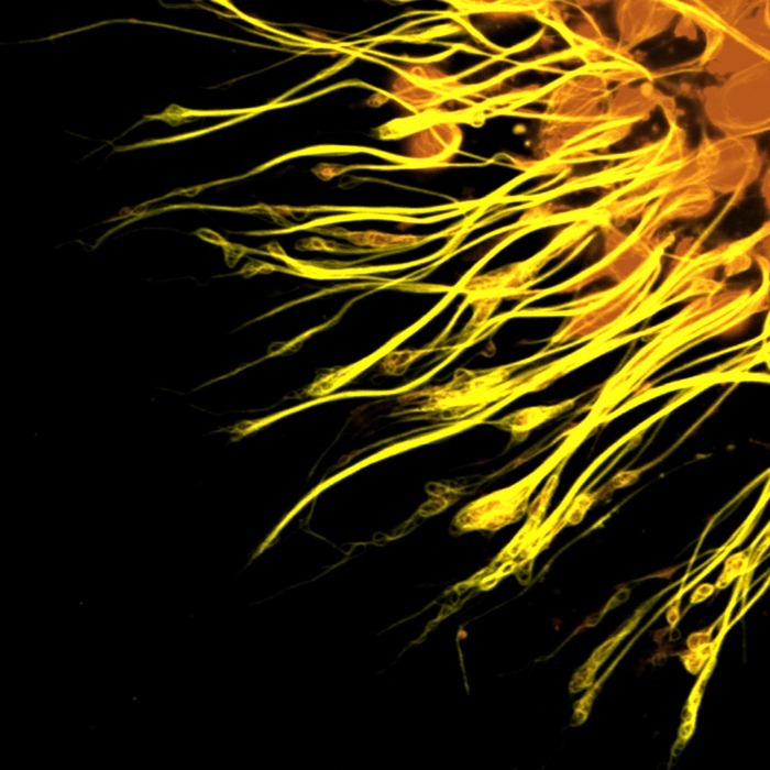
Photomicrograph of the molecular scaffolding of axons.

Photomicrograph of different components of the rat cerebellum, including Purkinje neurons in green, glia (non-neuronal cells) in red, and cell nuclei in blue.

Diffusion MRI image of a patient who has suffered a stroke in the thalamus. This has resulted in major disruptions to certain axon tracts, some of which are visible at the bottom of the figure.
[via PDN]
No comments:
Post a Comment